Root Canal Xray: Purpose, Procedure and Benefits

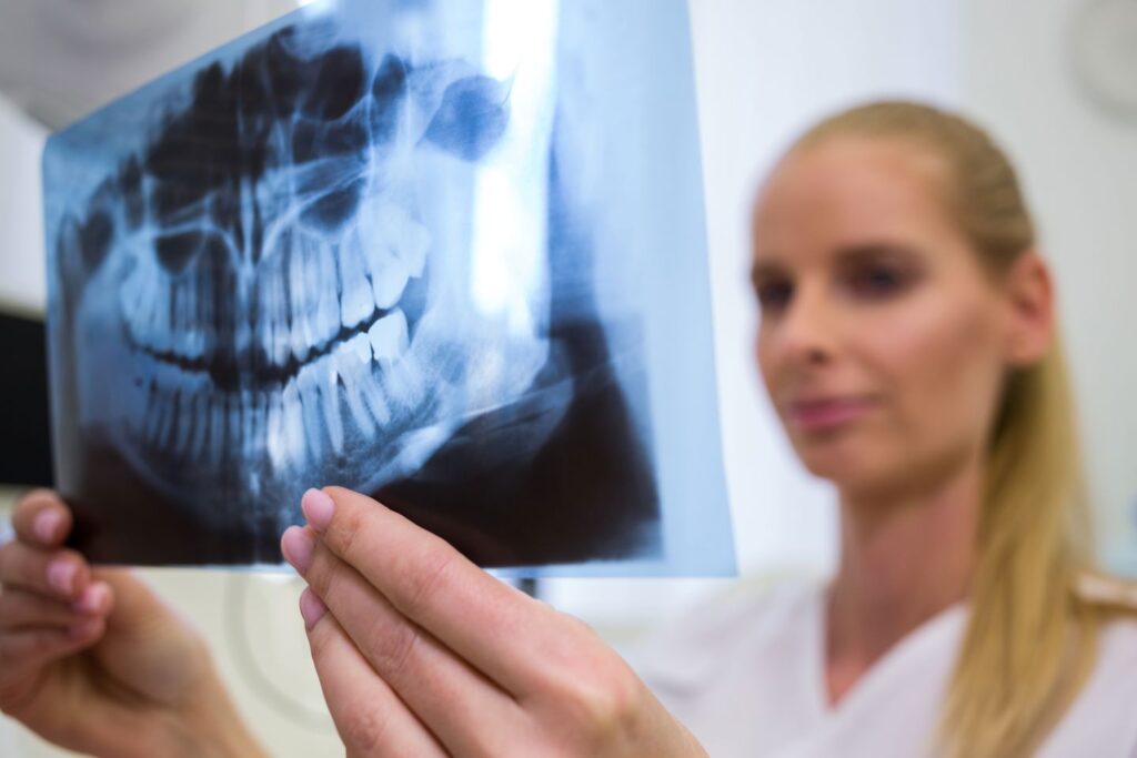
Root Canal Xray (also called periapical x-rays) have become an essential element of modern dentistry, serving as diagnostic tools that assess the health of tooth roots and surrounding structures. Here, we explore their purpose, procedure, benefits and any possible risks involved with root canal X-rays
Root Canal Xray Purpose
Root Canal X-rays serve the primary function of providing a detailed visual representation of one or more teeth or groups of them, which allows dentists to detect and diagnose various issues related to root canals affecting each root, including infections, decay, fractures and abnormalities. Furthermore, Root Canal xray offers unique insights into both the internal structures of each tooth and its surrounding bone.
Procedure for Root Canal X-ray
Root Canal Xrays are an integral component of dental diagnosis. Here’s a step-by-step overview:
Prep Work: Before commencing an X-ray examination, dental professionals will ensure your safety by providing a lead apron to safeguard against radiation exposure. This precautionary measure ensures the highest standards of patient well-being.
Optional Bitewing X-ray (optional): Dentists may begin with a bitewing X-ray, which captures images of both upper and lower teeth in one location. Bitewing X-rays allow dentists to readily identify cavities between teeth and gain a comprehensive view of dental conditions.
Periapical X-ray: At the core of this procedure lies periapical X-ray, wherein an X-ray machine is positioned to capture images of one tooth at a time – from crown tip to root tip and surrounding bone structure.
Post-Treatment X-ray: After conducting a root canal procedure, dentists may perform a follow-up Xray to assess its success and ensure the infection has been eliminated and healing has proceeded according to plan. These post-treatment X-rays allow dentists to stop the disease while ensuring your tooth heals as expected.
Benefits of Root Canal X-ray
Root Canal Xray offer many advantages to dental practitioners. Here are the main benefits:
Root Canal X-rays are Accurate Diagnosis Tools: One of the main advantages of root canal X-rays is their ability to provide accurate diagnostic information, enabling dentists to pinpoint the source of any dental issues for treatment purposes. Root Canal X-rays help dentists discover why patients experience dental discomfort, helping them create tailored strategies.
Treatment Planning: Root Canal Xray provides dentists with invaluable images for successfully planning and performing root canal treatments. Dentists can visualize the inner structures of each tooth, identify how many canals exist within the tooth, and anticipate any obstacles they might face during treatment.
Prevention: By early diagnosis of dental problems, root canal X-ray contribute to preventing complications. Early identification allows swift intervention to minimize further damage to the tooth and surrounding tissues.
Monitoring Healing: Post-treatment X-rays can provide dentists with invaluable data on the success of root canal treatments, ensuring that any infection has been eradicated and that teeth are healing as planned.
Patient Education: Root Canal Xray serves an educational function, allowing dentists to explain the nature of dental issues to patients and visualize affected areas to make treatment decisions more quickly and promote active participation in oral health.
Potential Concerns and Safety Measures
Root Canal Xray are generally safe; however, it’s essential that any potential issues be discussed and safety measures highlighted:
Radiation Exposure: While X-ray involves small doses of radiation, modern dental X-ray machines are designed to minimize exposure. Lead aprons and high-speed films help further reduce radiation risks.
Pregnancy and X-Ray: Individuals who are pregnant may worry about radiation exposure during gestation. To protect themselves effectively from radiation exposure, lead aprons offer adequate protection. Furthermore, dentists generally avoid unnecessary X-rays during the first trimester of gestation.
Alternative Imaging Technologies: When considering alternative imaging technologies such as cone-beam computed tomography (CBCT), three-dimensional images may provide more comprehensive views of dental structures and require less radiation exposure than traditional radiography methods. CBCT imaging can offer three-dimensional opinions that make for more thorough cases than two-dimensional radiographs alone.

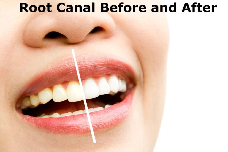
Root Canal Xray Before and After
While I cannot provide exact images, I can describe what an X-ray of the root canal will look like before or after treatment.
Root Canal Xray Before:
- Decay or Infection: In the initial scan, there may be apparent indications of decay or infection on the affected tooth. It may be visible as an eerie dark spot at the end of the tooth, signalling an infection within the canals of the root.
- Abscess or swelling: If an infection has grown, there could be signs of swelling or an abscess in the tooth, which can be seen as a radiolucent (dark) part of an X-ray.
- Inflammation of the surrounding tissues: The X-ray may be able to detect signs of inflammation within the bone or tissues surrounding it that indicate an intervention needs to be performed.
Root Canal Xray After:
- Cleared infection If the root is removed canal successfully, the X-ray post-treatment should show a decrease or absence of the previously diagnosed infection or decay. The dark zone surrounding the root’s tip must be reduced or eliminated.
- Well-Obturated Canal The X-ray will show a well-obturated (filled) canal for the root, and the filling material extended to the top of the root. This will ensure a good seal, lowering the infection risk recurring.
- Reduced Abscess or Swelling: If there was an abscess or swelling before the root canal, the post-treatment X-ray will reveal a decrease or resolution of these problems.
- Healing of the Surrounding tissues: Signs of healing, such as decreased inflammation within the bone and surrounding tissues, can be seen through X-rays.
- Stable Tooth Structure The X-ray must show an established tooth structure that is free of any indication of fractures or problems. The tooth being treated should be able to blend well with the dental anatomy surrounding it.
Bad Root Canal Xray
“Bad” root canal X-ray could include any problems visible on a radiograph that indicate potential issues with either the tooth or the root canal treatment. Some possible signs that your root canal X-ray is less-than-ideal include:
An Incomplete Root Canal Filling: If an X-ray reveals that the filling doesn’t reach the tip of the tooth root, this could indicate a preliminary treatment and leave bacteria room to continue their presence, potentially leading to persistent or recurrent infection.
Overfilled or Underfilled Canal: An X-ray may reveal problems with the amount of filling material used in root canal filling procedures. Excessive or inadequate filling may cause inflammation or infection to surrounding tissues and lead to complications that require medical intervention, including inflammation or infection of surrounding areas.
Perforation: If an X-ray reveals that a dentist created an unexpected hole during treatment, known as perforations, this may constitute a complication and require additional treatments to seal off and prevent future issues.
Missed Canal: To accurately assess root canal health and structure, an X-ray should show each canal’s number and shape. Missing one during treatment could result in persistent infection that necessitates additional interventions.
Poorly Obturated Canal: An X-ray may reveal deficiencies in the root canal filling material quality, such as leakage. If bacteria reenters the canal through holes left open by improper sealing materials, reinfection could occur and be an added source of pain and infection.
Apical Lesion or Cyst: On an X-ray, any radiolucency near the tip of the tooth’s root could indicate the presence of an apical lesion or cyst and could indicate continued infection or complications after root canal treatment.
Root Fracture: Though fractured roots may not always be visible on an X-ray, signs such as suspicious lines or dark shadows could signal possible fractures that compromise successful root canal treatment. Fractures could also result in additional medical problems.

Infected Root Canal Xray
An infected root canal X-ray typically exhibits sure indicative signs that indicate infection or inflammation at or around the roots of teeth. Here are some features that might be visible on such an X-ray:
Periapical Radiolucency: On an X-ray, there may be a dark area around the tip of a tooth’s root known as periapical radiolucency, which indicates bone density loss and could indicate inflammation or infection.
Widened Periodontal Ligament Space: When infected with bacteria, the periodontal ligament space – typically appearing as a thin dark line around the root of healthy teeth – may widen due to inflammation and bone loss in surrounding tissues.
Loss of Tooth Structure: If infection has led to tooth decay or destruction, its effects may become apparent on an X-ray image in the form of dark areas or changes in the shape and size of teeth.
Swelling or Abscess Formation: In severe cases, an X-ray might show signs of swelling or the development of an abscess – represented as a radiolucent area around a tooth root, which indicates an accumulation of pus.
Pulpal Changes: An X-ray may reveal changes in the pulp chamber and root canal system, including irregularities or widening of canal space due to bacteria infection and inflammation processes in the pulp.
Important Note: These features may differ depending on the severity and duration of an infection and individual patient factors. A dentist or endodontist would utilize these radiographic findings in combination with clinical examination results and patient symptoms to diagnose and plan treatment plans for an infected root canal.
Root Canal Shadow on X-Ray
Root Canal Shadow on X-Ray On an X-ray dental image, “shadows” can indicate various findings; when applied to root canal treatments, these shadows could represent anything from different findings, such as fractured roots to material leakage from the inside. Here are some interpretations for possible interpretations of shadowed images:
Normal Root Canal Filling Shadow:
Following a successful root canal procedure, post-treatment X-ray should show a shadow along the length of each treated root canal, indicating filling material within. This is considered normal.
Root Canal Filling Shadow: Inadequate Root Canal Filling Procedures.
If the root canal filling appears incomplete or inadequate on an X-ray, this could indicate that treatment hasn’t reached all parts of the canal and left space for bacteria to persist, potentially leading to ongoing or recurrent infection.
Periapical Radiolucency Shadow:
Dark areas or radiolucent spots around the root tip can indicate periapical pathology, such as abscesses or inflammation. These dark areas suggest something is amiss at its tip, perhaps an infection or unresolved complications.
Shadow Suggests Fracture:
Root fractures can be complex to diagnose; they typically appear as dark lines or irregular shadows on an X-ray. Understanding dental X-rays requires professional expertise; only an experienced dentist or endodontist can accurately interpret a root canal X-ray and address potential issues within its borders.
Conclusion
Root Canal Xray is indispensable in modern dentistry, helping dentists diagnose and treat dental issues precisely. Their detailed images assist dentists in making accurate diagnoses, planning effective treatment, and monitoring the healing process. While radiation exposure concerns exist, safety measures like lead aprons and modern X-ray machines help ensure that benefits outweigh the risks, with technology continuously progressing into dental imaging tools to aid in optimal patient care.